

Biophysical-characterization-of-cell-cohesion
Volker Spindler
Franziska Vielmuth
Jens Waschke
Andrea Wehmeyer
We use a set of biophysical and imaging techniques to gain mechanistic insight into the function of cell adhesion molecules in cellular context. We here focus on the desmosome, an intercellular adhesive complex that confers strong cell cohesion to epithelial and some non-epithelial tissues (see here). The transmembrane adhesion molecules in the desmosome, the desmogleins and desmocollins, are also located extradesmosomal in the membrane. The function and binding properties of these molecules are largely unknown. Furthermore, it is unclear how the adhesive properties and the localization of desmosomal adhesion molecules in the membrane are regulated by intracellular signaling cues and are impaired in disease models (pemphigus, arrhythmogenic cardiomyopathy, Crohn’s disease).
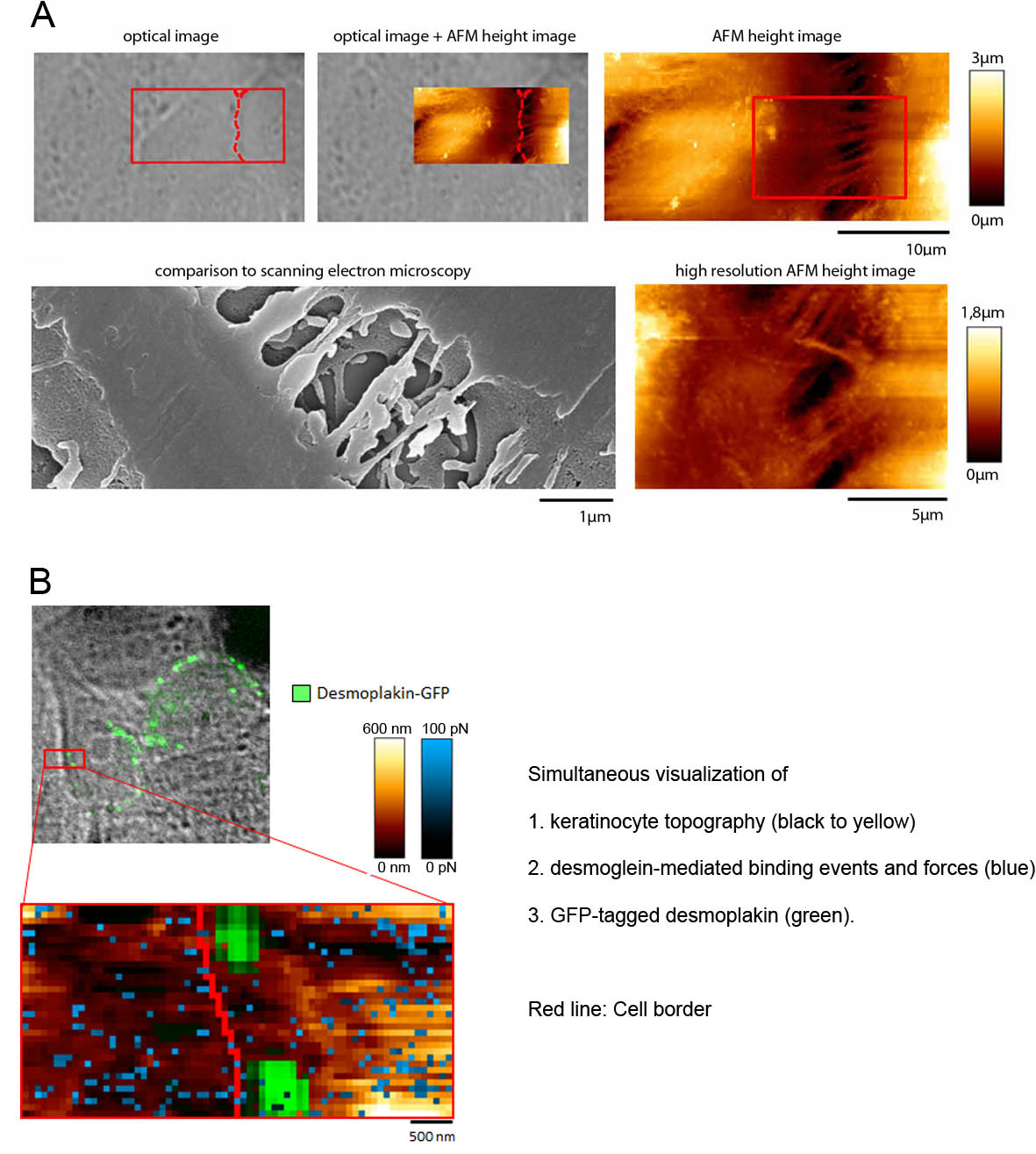
To address these questions, besides standard biochemical and molecular biology techniques, we apply atomic force microscopy, which enables us to delineate the topography of a cell and simultaneously to quantify the distribution, binding forces and additional binding properties of a specific cell adhesion molecule (Fig. 1).
In another biophysical approach, we use optical tweezers which allow the investigation of cell surface-attached microbeads that were functionalized with specific adhesion molecules. Binding properties of these beads can be determined by exerting forces using laser beams.
Recent publications (2013 to present):
Spindler V, Meir M, Vigh B, Flemming S, Hütz K, Germer CT, Waschke J, Schlegel N (2015). Loss of desmoglein2 contributes to the pathogenesis of Crohn’s disease. Inflamm Bowel Dis, in press
Flemming S, Burkard N, Renschler M, Vielmuth F, Meir M, Schick MA, Wunder C, Germer CT, Spindler V, Waschke J, Schlegel N (2015). Soluble VE-cadherin is involved in endothelial barrier breakdown in systemic inflammation and sepsis. Cardiovasc Res, ePub: DOI: 10.1093/cvr/cvv144
Vielmuth F, Hartlieb E, Kugelmann D, Waschke J, Spindler V (2014); Atomic force microscopy identifies regions of distinct desmoglein 3 adhesive properties on living keratinocytes; Nanomedicine, 11(3):511-20
Monteiro AC, Luissint AC, Sumagin R, Lai C, Vielmuth F, Wolf MF, Laur O, Reiss K, Spindler V, Stehle T, Dermody TS, Nusrat A, Parkos CA (2014). Trans-dimerization of JAM-A Regulates Rap2 and is Mediated by a Domain that is Distinct from the Cis-dimerization Interface; Mol Biol Cell, 25(10):1574-85
Spindler V, Rötzer V, Dehner C, Kempf B, Gliem M, Radeva M, Hartlieb E, Harms GS, Schmidt E, Waschke J (2013). Peptide-mediated desmoglein 3 crosslinking prevents pemphigus vulgaris autoantibody-induced skin blistering; J Clin Invest, 123(2):800–811
Hartlieb E, Partilla M, Vigh B, Spindler V, Waschke J (2013). Desmoglein 2 is less important than Dsg3 for keratinocyte cohesion PLOS One, 8(1): e53739