

Epidermal cohesion and pemphigus (together with Jens Waschke)
Jens Waschke and Volker Spindler (group leaders),
Eva Arnold (postdoctoral fellow)
Franziska Vielmuth (postdoctoral fellow),
Vera Rötzer (PhD student)
Desalegn Egu (PhD student)
Elias Walter (MD student)
Julia Winkler (MD student),
Martina Hitzenbichler (technician)
Sabine Mühlsimer (technician)
Andrea Wehmeyer (technician)
The epidermis represents the outermost barrier sealing our body against the environment. The integrity of these stratifying cell layers depends on desmosomes, specific intercellular junction complexes designed to tether adjacent keratinocytes together. This enables the epidermis to withstand the high mechanical forces this tissue constantly is exposed to. The core of desmosomes is composed of the cadherin-type transmembrane adhesion molecules desmogleins and desmocollins. These molecules are intracellularly linked to the intermediate filament network by the adapter proteins plakoglobin, plakophilins and desmoplakin (Fig. 1).

Fig. 1 Schematic of a desmosome
Specifically desmogleins are known to exist “free” on the membrane without attachment in the desmosome. These molecules are connected to a variety of signaling molecules such as Rho-GTPases, Src and p38MAPK. By regulating the activity of these molecules, extradesmosomal desmogleins serve as signaling hubs to modulate cellular behavior (Fig. 2).
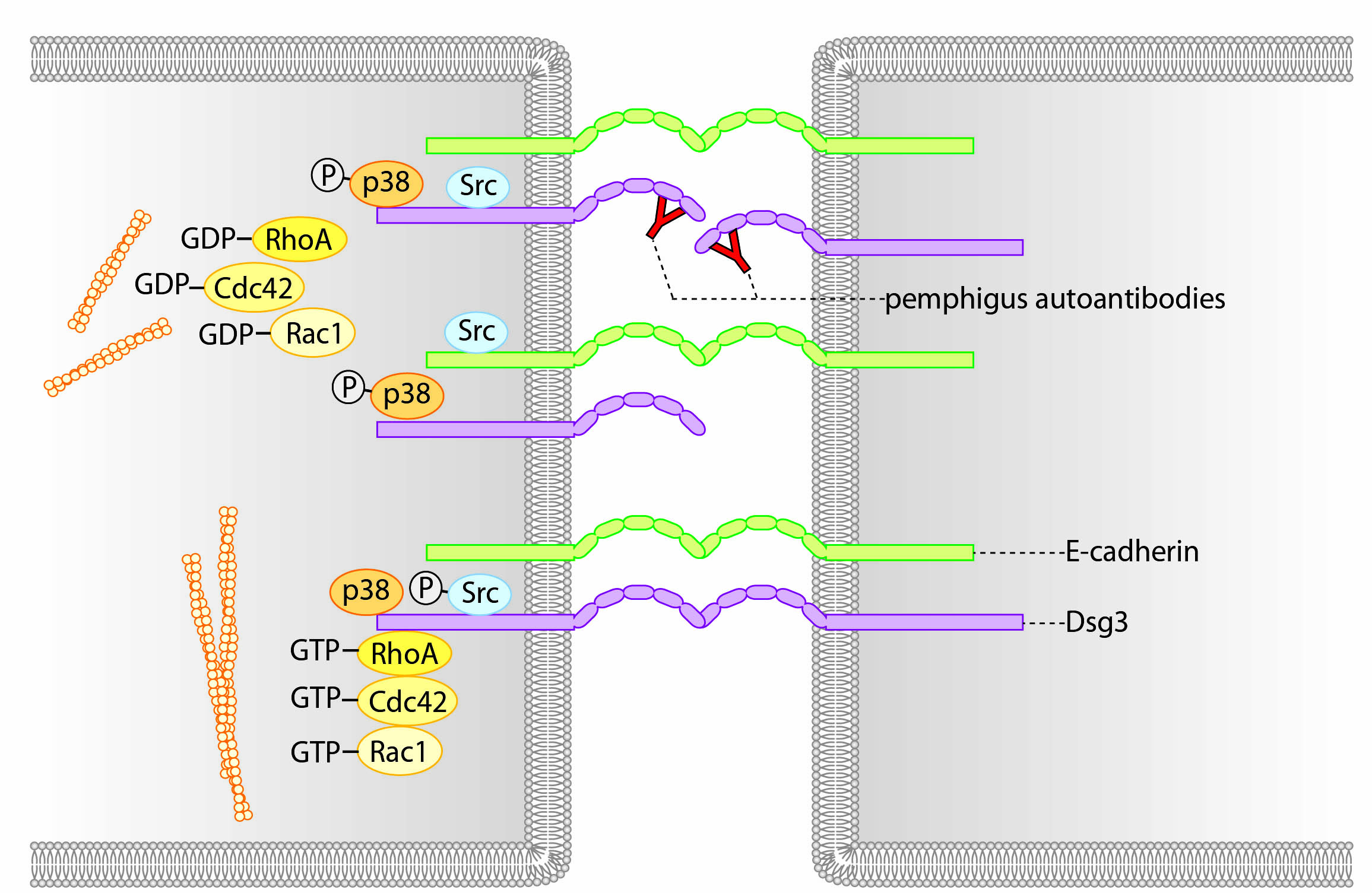
Fig. 2 Extradesmosomal desmoglein molecules serve as signaling hubs
The importance of desmosomal adhesion becomes evident by the autoimmune disease pemphigus. The main clinical variant is pemphigus vulgaris. Patients develop autoantibodies against the desmoglein isoforms 3 and 1 (Dsg3 and Dsg1) resulting in loss of desmosomal function. This loss of cell cohesion leads to cleft formation within the epidermis and the mucosa of the oral cavity. Macroscopically, blisters are visible which rupture rapidly (Fig.3). Without immunosuppression, the disease is lethal.
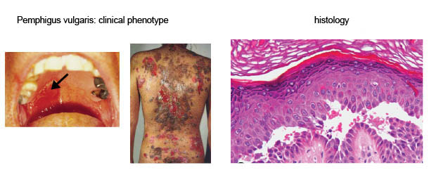
Fig. 3: Pemphigus
Binding of pemphigus autoantibodies leads to altered intracellular signaling, at least in part following initial loss of Dsg3 interaction (Fig. 4). This induces (i) loss of Dsg3 and other desmosomal molecules from the cell surface and (ii) uncoupling of the intermediate filaments from the desmosome. These two effects are thought to represent the main mechanisms of loss of cell-cell adhesion and ultimately blister formation.
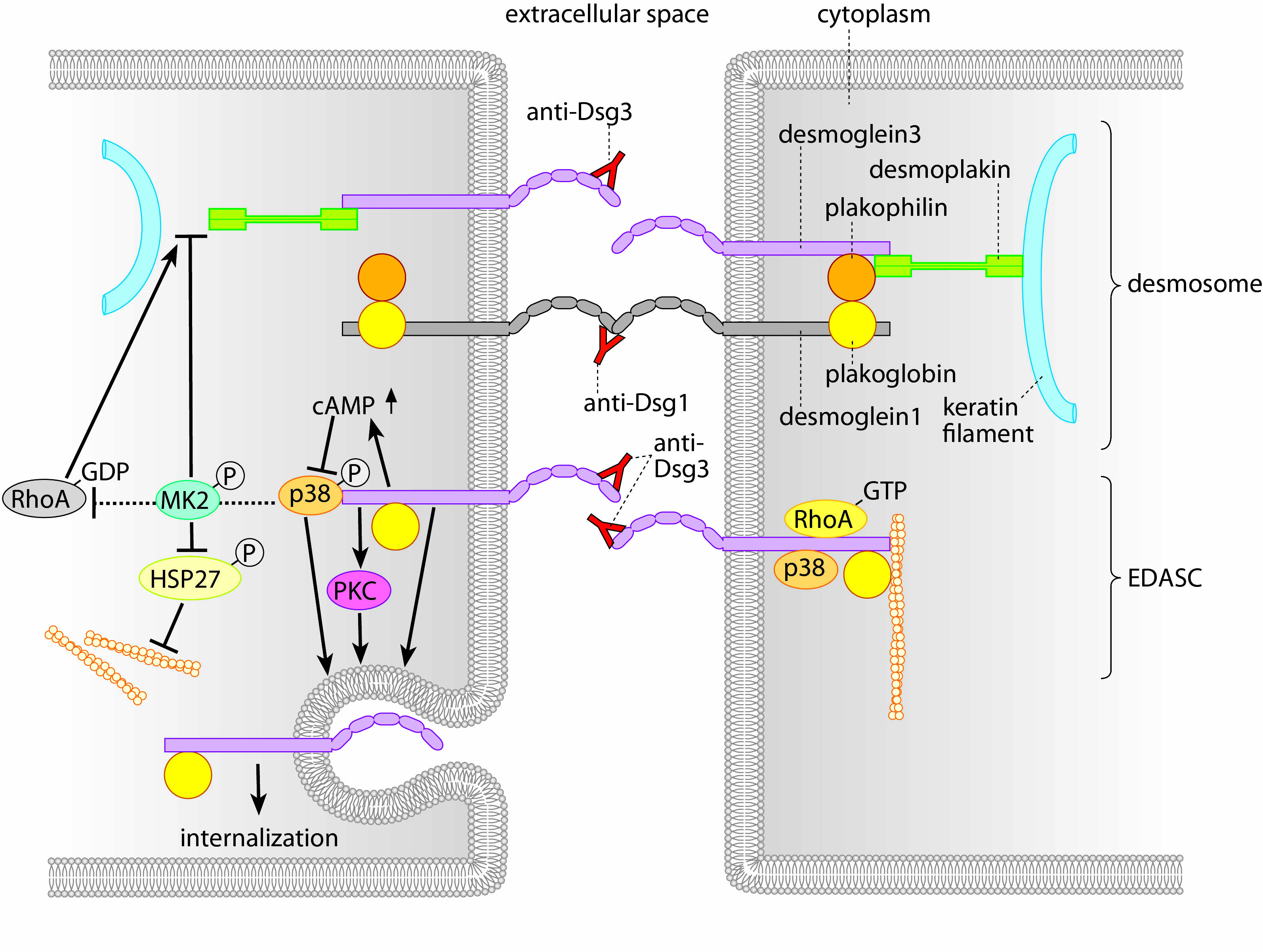
Fig. 4: Mechanisms of blister formation in pemphigus
Within the project we investigate the regulation of desmosomal adhesion. Specifically, we address how desmosomes are assembled and disassembled and which mechanisms and signals are involved in these processes. Furthermore, we aim to better understand the mechanisms that lead to loss of cell cohesion and blister formation in pemphigus to identify novel targets as potential treatment options for this life-threatening disease.
We combine state-of-the-art biochemical and molecular biology techniques with morphological assessment of desmosomes and their turnover (using live cell imaging, confocal microscopy, transmission and scanning electron microscopy). To correlate this with the adhesive function of desmosomes, we apply adhesion assays and biophysical techniques such as atomic force microscopy and optical tweezers (see here).
Recent publications (2013 to present):
Dehner C, Rötzer V, Waschke J, Spindler V (2014). A desmoplakin point mutation with enhanced keratin association ameliorates pemphigus vulgaris autoantibody-mediated loss of cell cohesion. Am J Pathol, 184(9):2528-36.
Hartlieb E, Rötzer V, Radeva M, Spindler V, Waschke J (2014). Desmoglein 2 compensates for desmoglein 3 but does not control cell adhesion via regulation of p38-mitogen-activated protein-kinase in keratinocytes. J Biol Chem, 289(24):17043-17053
Rötzer V, Breit A, Waschke J, Spindler V (2014). Adducin is required for keratinocyte cohesion . J Biol Chem, 289(21):14925-40
Spindler V, Dehner C, Hübner S, Waschke J (2014). Plakoglobin but not desmoplakin regulates keratinoycte cohesion via p38MAPK signaling. J Invest Dermatol, 134(6):1655-64
Spindler V, Rötzer V, Dehner C, Kempf B, Gliem M, Radeva M, Hartlieb E, Harms GS, Schmidt E, Waschke J (2013). Peptide-mediated desmoglein 3 crosslinking prevents pemphigus vulgaris autoantibody-induced skin blistering; J Clin Invest, 123(2):800–811
Hartlieb E, Partilla M, Vigh B, Spindler V, Waschke J (2013). Desmoglein 2 is less important than Dsg3 for keratinocyte cohesion PLOS One, 8(1): e53739