

Meibomian gland cells and Dry eye syndrom
Members
Vera Rötzer and Jens Waschke (group leaders)
Francesca Melega (MD student)
Martina Hitzenbichler (technician)
In our new research group, we focus on the cell adhesion in Meibomian glands and their role in the development of the Dry eye syndrome. Meibomian glands are modified, holocrine sebaceous glands within the upper and lower eyelids essential for the maintenance of the integrity and the health of the ocular surface. During movement from the basal (proliferating) to the central zone of the acini, specialized Meibomian gland epithelial cells (meibocytes) mature and synthesize lipids, which spread onto the tear film and thus prevent the evaporation of the cornea. In this process of maturation and differentiation, meibocytes display a characteristic morphology, e.g. loss of nuclei, and a significantly increased frequency of desmosomes (Fig.1).
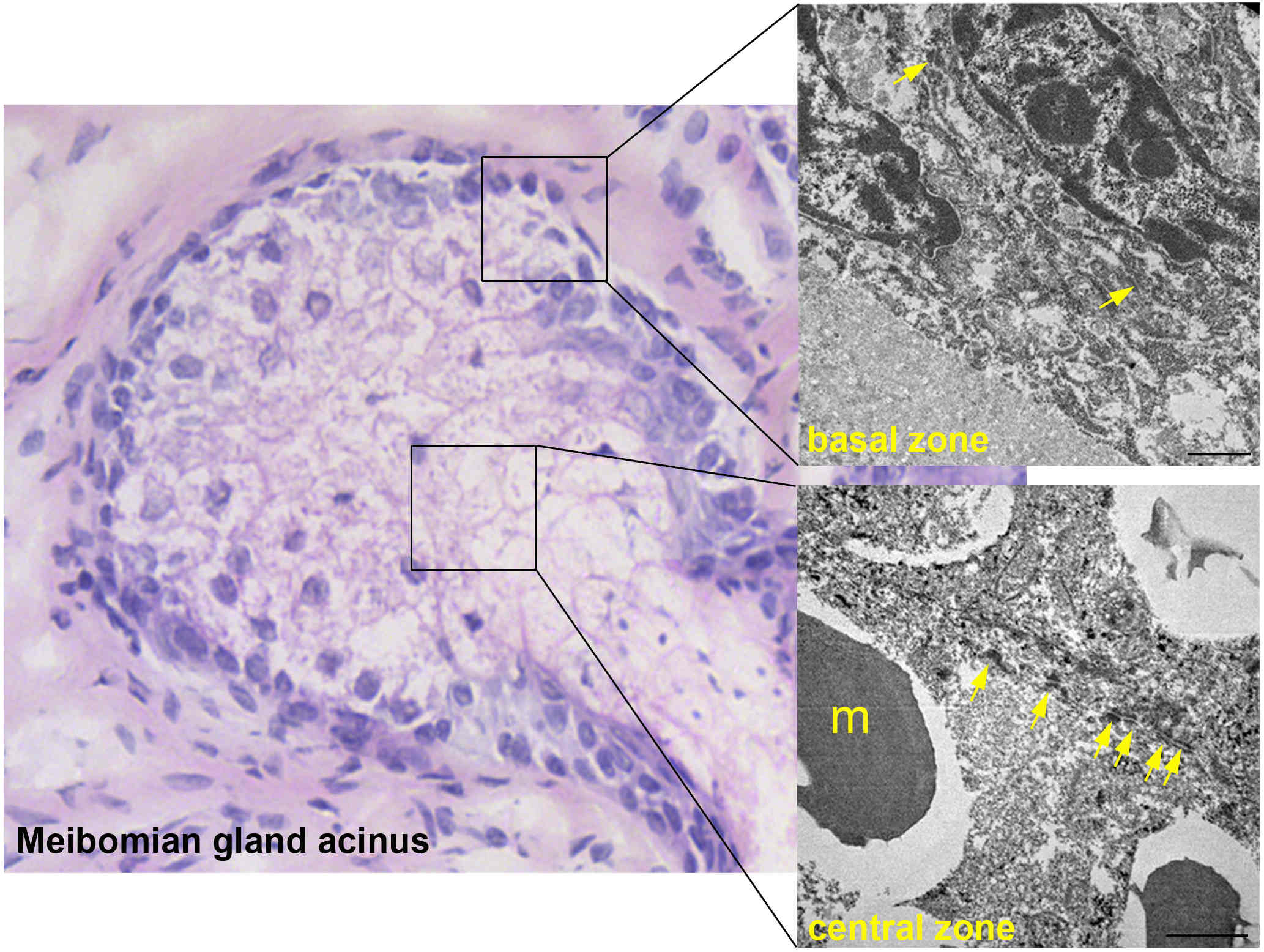
Figure 1: Differentiation-dependent distribution of desmosomes within Meibomian gland acinus
Desmosomes are normally abundant in tissues exposed to mechanical stress such as the epidermis of the skin or the myocardium and provide strong intercellular cohesion. The clinical relevance of intact intercellular contacts is underscored by diseases like pemphigus vulgaris or arrythmogenic cardiomyopathy.
The impact of intercellular cohesion between meibocytes has not been investigated so far but might be of relevance since the Dry eye syndrome is part of the PV pathogenesis. The Dry eye syndrome, from which more than 20% of the European populations suffers, is strongly associated with the dysfunction of Meibomian glands (MGD). Therefore, with our research we address intercellular adhesion of Meibomian gland cells and all accompanying signaling pathways.
Beside the correlation between lipid synthesis and the frequency of desmosomes, meibocytes display a specific, differentiation-dependent pattern of adhesion molecules (Fig. 2, shown for desmosomal proteins Dsg1, Dsg2, Dp and for the adherens junction protein E-cadherin).
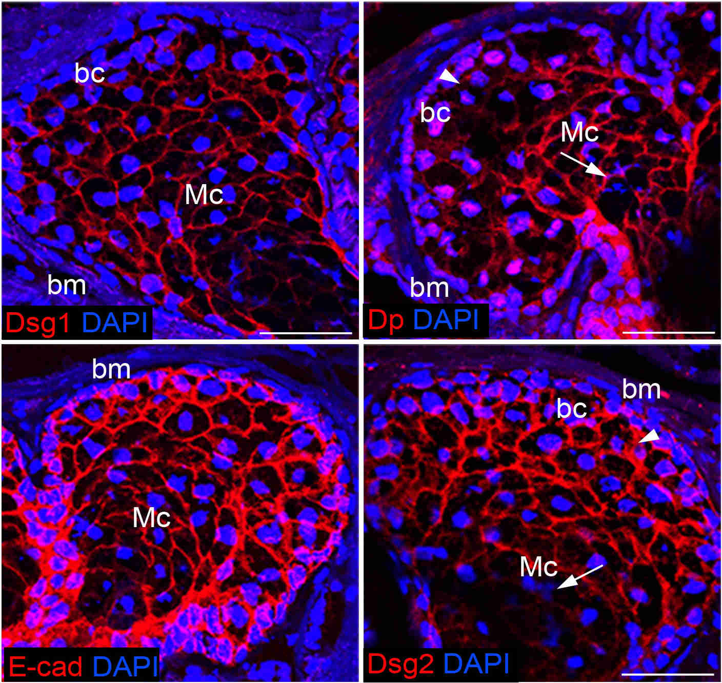
Figure 2: Differentiation-dependent distribution of adhesion molecules within Meibomian gland acinus
Recent publications (2015 to present)
Rötzer V, Egu D, Waschke J. (2016) Meibomian gland cells display a differentiation-dependent composition of desmosomes. Histochemistry Cell Biolology. 146(6)
Vielmuth F, Rötzer V, Hartlieb E, Hirneiß C, Waschke J, Spindler V. (2015) Pemphigus autoantibodies induce blistering in human conjunctiva. Investigative Ophthalmology & Visual Science (IOVS). 57(10)